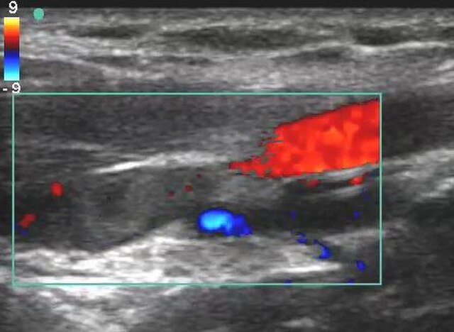
Venous Doppler ultrasound is a non-invasive diagnostic tool used to assess the health of the veins and blood flow within the body. It uses sound waves to create images of blood flow in veins, allowing doctors to diagnose a variety of venous disorders, such as deep vein thrombosis (DVT), varicose veins, and venous insufficiency. In this blog post, we’ll dive into what venous Doppler is, how it works, its uses, and what you can expect during the procedure.
What is a Venous Doppler Ultrasound?
A venous Doppler ultrasound is a type of imaging test that uses high-frequency sound waves to examine blood flow in the veins. Unlike X-rays or CT scans, which use radiation, Doppler ultrasound is completely safe and painless. The test helps to visualize how well blood flows through the veins, especially in areas like the legs, thigh and groin, where venous problems are common.
The term “Doppler” refers to the Doppler effect, which describes how sound waves change frequency when they bounce off moving objects. In the case of a venous Doppler, the "moving objects" are red blood cells. By detecting the movement of these cells, the Doppler technology can assess the speed and direction of blood flow.
How Does a Venous Doppler Work?
During a venous Doppler ultrasound, a special gel is applied to the skin, and a small handheld device called a transducer or probe is used. The transducer sends high-frequency sound waves into the body, which bounce back off blood cells moving through veins. These sound waves are then converted into images that display blood flow, allowing the technician or physician to assess the condition of the veins.
In addition to imaging, the Doppler ultrasound can also measure the speed of blood flow and check for irregularities such as blockages, blood clots, or vein insufficiency. It is often used to assess veins in the legs, as this is the area where venous problems are most prevalent.
Common Uses of Venous Doppler
Venous Doppler ultrasound is typically used to diagnose or monitor various venous conditions, including:
1. Deep Vein Thrombosis (DVT)
- DVT occurs when a blood clot forms in a deep vein, often in the legs. These clots can be dangerous if they break loose and travel to the lungs (pulmonary embolism). Venous Doppler is the gold standard for diagnosing DVT as it can visualize the clot and assess whether it is blocking blood flow.
2. Venous Insufficiency
- This occurs when veins, especially in the legs, are unable to return blood effectively to the heart. Venous insufficiency can lead to swelling, pain, and varicose veins. A venous Doppler can identify areas of poor blood flow or valve dysfunction in veins.
3. Varicose Veins
- Varicose veins are enlarged, twisted veins that often appear in the legs. Venous Doppler can help evaluate the severity of varicose veins and determine if there is any underlying venous insufficiency causing them.
4. Chronic Venous Disease
- Chronic venous disease (CVD) refers to long-term venous issues that lead to symptoms like leg pain, swelling, and skin changes. Venous Doppler ultrasound can help assess the condition of the veins and assist in treatment planning.
Why is Venous Doppler Important?
Venous Doppler ultrasound is crucial because it helps diagnose venous disorders early, often before symptoms become severe. Early detection can lead to more effective treatments and prevent complications such as chronic pain, ulcers, or even life-threatening conditions like pulmonary embolism.
Here are a few reasons why venous Doppler is important:
- Non-invasive: The procedure does not require incisions, needles, or radiation, making it a safe and comfortable option for most patients.
- Accurate Diagnosis: Doppler ultrasound provides real-time images of blood flow, enabling doctors to diagnose conditions like blood clots or varicose veins with high accuracy.
- Guides Treatment: Whether it's medication, lifestyle changes, or surgery, venous Doppler helps guide treatment plans by providing detailed insights into the condition of the veins.
- Prevention of Complications: Identifying venous issues early can help prevent more serious conditions, such as chronic venous insufficiency or pulmonary embolism.
What to Expect During a Venous Doppler Ultrasound
A venous Doppler ultrasound is typically performed in a doctor's office or a diagnostic imaging center. The procedure is quick, non-invasive, and usually takes around 20-30 minutes.
Here’s what you can expect:
- Preparation: You may be asked to remove clothing or jewelry from the area being examined (usually the legs). You’ll be provided with a gown or cover sheets for privacy.
- The Procedure: The operator will apply a cool gel to the skin over the area of interest, such as the legs. The gel helps the transducer make better contact with your skin. The operator will move the transducer over your skin to capture images and assess blood flow.
- Duration: The test is relatively quick, typically lasting 20 to 30 minutes, depending on the area being examined.
- After the Procedure: There is no downtime after a venous Doppler ultrasound. You can resume your normal activities immediately, and the results will be reviewed by your doctor to determine the next steps in your care.
Conclusion
Venous Doppler ultrasound is a valuable, non-invasive tool for diagnosing and monitoring venous conditions. Whether you’re dealing with varicose veins, chronic leg pain, or a suspected blood clot, this test provides essential insights into your vascular health. If you’re experiencing symptoms of venous issues, such as swelling, pain, or visible veins, consult your doctor, who may recommend a venous Doppler to better understand your condition and guide effective treatment.
