The anomaly scan, also known as the mid-pregnancy ultrasound or detailed fetal scan, is usually performed between 18 and 22 weeks of pregnancy. This scan plays a crucial role in assessing the structural development of the baby and identifying potential abnormalities that may require further investigation, monitoring, or management.
Below are the top 10 abnormalities commonly detected during an anomaly scan:
1. Neural Tube Defects (e.g., Spina Bifida)
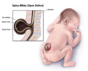
These are defects of the brain, spine, or spinal cord. Spina bifida is one of the most serious and detectable neural tube defects, where the spinal column doesn’t close completely. The scan may reveal an open spinal defect, abnormal head shape ("lemon sign"), or fluid buildup in the brain ("banana sign").
2. Cleft Lip and/or Palate
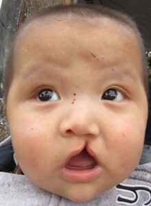
A cleft lip can usually be detected during the anomaly scan. It appears as a separation or split in the upper lip, which may be unilateral or bilateral. A cleft palate is harder to detect but may be suspected if other facial abnormalities are present.
3. Congenital Heart Defects
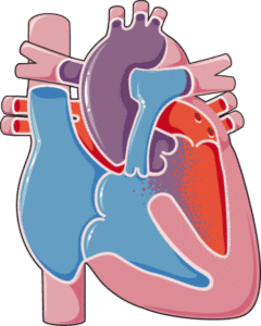
Structural problems with the heart, such as hypoplastic left heart syndrome, ventricular septal defects (VSD), or transposition of the great arteries, are among the most common congenital anomalies. The sonographer examines the four-chamber view and outflow tracts of the fetal heart to identify these.
4. Limb Abnormalities
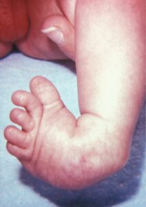
Abnormalities like shortened limbs (associated with skeletal dysplasias), missing limbs, or clubfoot can often be picked up during the scan. The sonographer checks all four limbs for length, symmetry, and position.
5. Abdominal Wall Defects
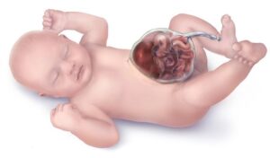
Conditions like gastroschisis (where the intestines are outside the body) and omphalocele (where abdominal organs protrude into the umbilical cord and are covered by a membrane) are visible on the scan. These defects may require surgical correction after birth.
6. Brain Abnormalities (e.g., Hydrocephalus)

The anomaly scan can detect ventriculomegaly (enlargement of the brain's fluid spaces), absence of the corpus callosum, or anencephaly (absence of a major portion of the brain and skull). These may indicate serious neurological concerns.
7. Renal (Kidney) Abnormalities
Issues like hydronephrosis (swelling of one or both kidneys due to urine buildup) or multicystic dysplastic kidney (where the kidney is replaced by cysts) are often detectable. These may be isolated or part of a syndrome.
8. Diaphragmatic Hernia
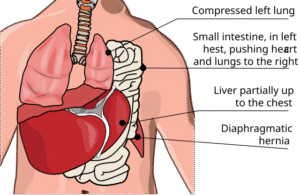
This condition occurs when abdominal organs like the stomach or intestines move into the chest cavity through a hole in the diaphragm. It can lead to underdeveloped lungs and may be life-threatening if severe.
9. Bladder Outlet Obstruction
A condition such as posterior urethral valves in male fetuses causes obstruction of urine flow, leading to an enlarged bladder and potential kidney damage. It’s detectable via a persistently full bladder and dilated urinary tract on ultrasound.
10. Chromosomal Abnormality Markers
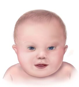
While not diagnostic, certain "soft markers" like nuchal thickening, echogenic bowel, shortened femur or humerus, and absent nasal bone may suggest an increased risk of chromosomal abnormalities such as Down syndrome (Trisomy 21), Edwards syndrome (Trisomy 18), or Patau syndrome (Trisomy 13).
Conclusion
Anomaly scans are vital for early detection of structural abnormalities, offering a window of opportunity for further diagnostic testing, parental counseling, and planning for medical or surgical interventions post-delivery if necessary. While not all abnormalities are detectable through ultrasound, many significant issues can be identified early, aiding in better outcomes for both mother and baby.