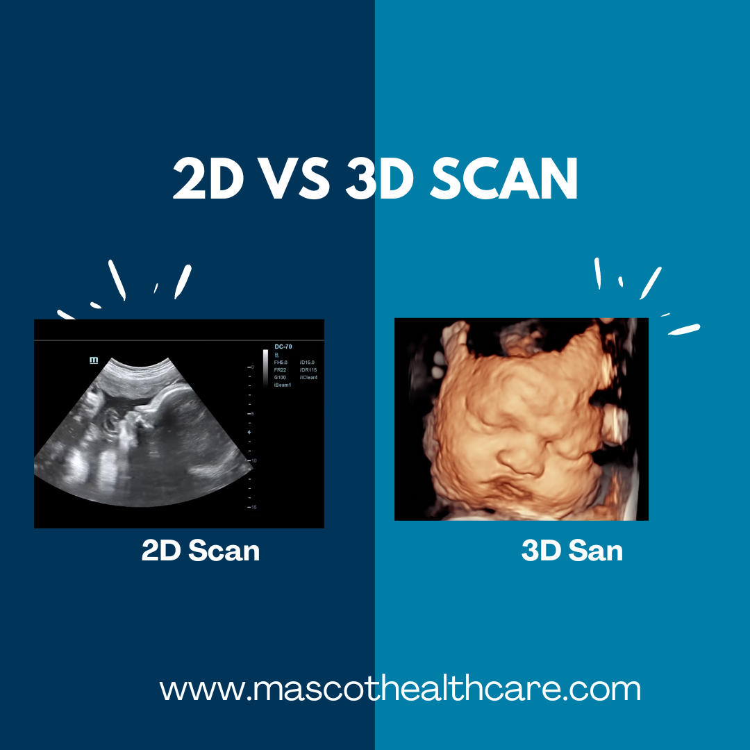
Q1: What is a 3D ultrasound?
A: A 3D ultrasound creates a three-dimensional image of the baby by combining multiple two-dimensional images taken at different angles. This method allows parents to see more detailed and realistic images of their baby's face and body, which can be particularly useful for identifying certain congenital anomalies and assessing fetal development more precisely.
Q2: What is a 4D ultrasound?
A: A 4D ultrasound takes the concept of 3D imaging a step further by adding the element of time, producing a live video effect. This allows parents to observe their baby's movements in real-time, such as yawning, stretching, or even smiling. The fourth dimension, time, adds an emotional component to the experience, making it a cherished memory for parents.
Q3: What are the benefits of 3D and 4D ultrasounds?
- Enhanced Diagnostic Capabilities: These ultrasounds provide more realistic detailed images, which can help in the detection of potential issues such as cleft lip, spinal problems, or heart defects. This allows for better planning and management of the pregnancy.
- Emotional Connection: Seeing a detailed image or live video of the baby can strengthen the emotional bond between parents and their unborn child. It provides a more tangible connection to the baby and can be a reassuring and joyful experience.
Q4: When are 3D and 4D ultrasounds typically performed?
A: These ultrasounds are typically performed between 24 and 32 weeks of pregnancy. This is when the baby has developed enough fat under the skin to provide clear images, but there is still enough amniotic fluid to get good pictures.
Q5: Are 3D and 4D ultrasounds safe?
A: Yes, 3D and 4D ultrasounds are considered safe when performed by a trained professional. They use the same type of sound waves as standard 2D ultrasounds, which have been used safely in obstetrics for decades.
Q6: Do I need a 3D or 4D ultrasound?
A: While 3D and 4D ultrasounds can provide additional details and a more emotional connection to the baby, they are not typically necessary for standard antenatal care. They are often considered optional and may not be covered by HMO. It’s best to discuss with your doctor to determine if these ultrasounds are necessary for your pregnancy.
Q7: How can I prepare for a 3D or 4D ultrasound?
A: Preparation is generally the same as for a 2D ultrasound. It’s helpful to stay hydrated in the days leading up to the ultrasound, as good hydration can improve the quality of the images. Follow any specific instructions provided by your doctor or the ultrasound operator.
Q8: What can I expect during the procedure?
A: During a 3D or 4D ultrasound, you will lie on an exam table, and a gel will be applied to your abdomen. The ultrasound technician will then use a transducer to send sound waves into your body, creating images of the baby on a screen. The procedure is non-invasive and typically takes about 10 to 45 minutes.
Q9: Can I get printed images or videos from the ultrasound?
A: Many facilities offer the option to get digital or hardcopy images or videos of the 3D or 4D ultrasound. This can be a wonderful keepsake for parents and family members.
Q10: When might facial images not be acquired during a 3D or 4D ultrasound?
A: Sometimes, it may not be possible to obtain clear facial images of the baby during a 3D or 4D ultrasound. This can occur due to several reasons:
- Baby's Position: If the baby is facing the mother's back, is turned sideways, or has their face pressed against the uterine wall, it can be difficult to get a clear image.
- Placenta Location: If the placenta is positioned in front of the baby's face, it can obstruct the view.
- Amniotic Fluid Levels: Adequate amniotic fluid is necessary for clear images. Low levels of amniotic fluid can make it challenging to get good pictures.
- Baby's Movements: Active movements or if the baby has hands, arms, or the umbilical cord covering their face can also prevent clear imaging.
In such cases, the ultrasound operator may ask the mother to change positions or return for a follow-up appointment to try again.
By understanding the benefits, process, and potential limitations of 3D and 4D ultrasounds, you can make informed decisions about your prenatal care and enjoy the unique experience of seeing your baby in remarkable detail before birth.
