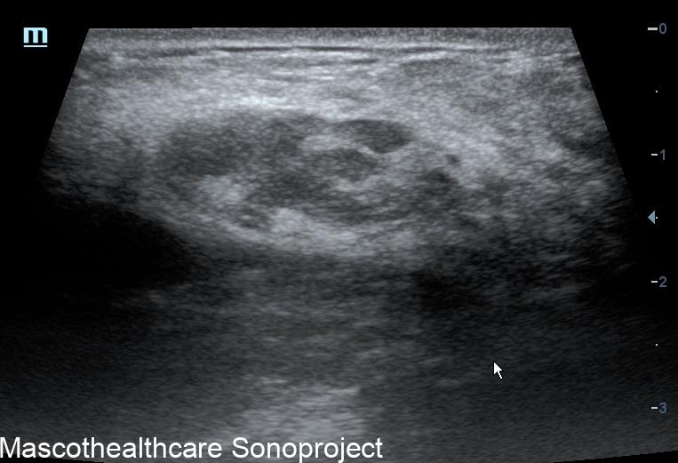
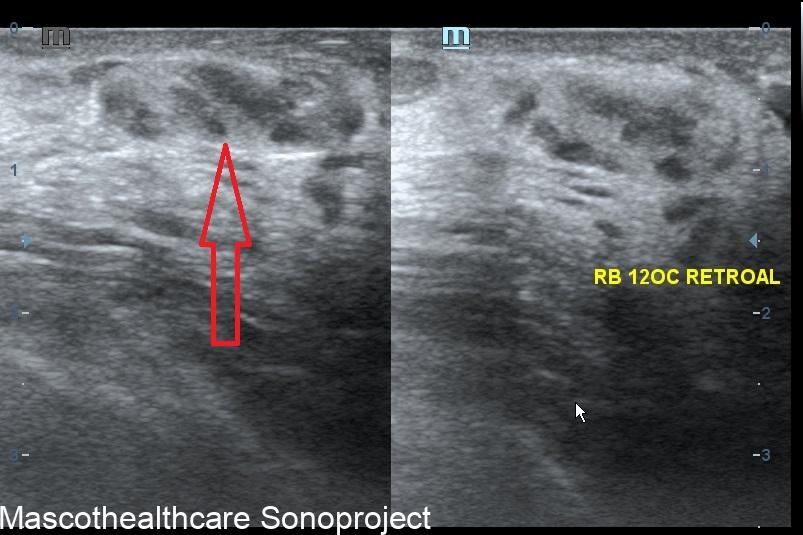
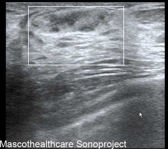
A breast hamartoma is a benign tumor-like lesion that consists of a disorganized mixture of normal breast tissues, including glandular, adipose (fatty), and fibrous tissues. These lesions are usually non-cancerous and do not increase the risk of breast cancer. Breast hamartomas are commonly found incidentally during routine breast imaging studies, such as mammography or ultrasound. Here are some key points about breast hamartomas:
- Incidence:
- Breast hamartomas are relatively common benign lesions and are often discovered incidentally during breast imaging studies.
- Age and Gender:
- They can occur at any age but are most frequently found in women between the ages of 35 and 55.
- Clinical Presentation:
- Most breast hamartomas are asymptomatic and are often discovered during routine breast screening. They typically present as painless, well-defined mass(es) within the breast.
- Imaging Characteristics:
- Mammography: On mammograms, breast hamartomas typically appear as well-circumscribed masses with a mixed density due to the combination of glandular and fatty tissues. They are described as the "breast within breast" appearance.
- Ultrasound: Ultrasound features of breast hamartomas include a heterogeneous echotexture due to the presence of various tissue types. They often have well-defined margins and may contain cystic spaces. The "breast within breast" appearance an also be elicited here.
- Histological Features:
- Under a microscope, breast hamartomas show a disorganized mixture of epithelial and stromal components, resembling normal breast tissues. The glandular elements may form duct-like structures, and the stromal component may include fibrous and adipose tissue.
- Differential Diagnosis:
- Breast hamartomas should be distinguished from other breast lesions, such as fibroadenomas, cysts, and malignant tumors. Typical imaging features of higher BIRADS lesion are almost always absent.
- Treatment:
- As breast hamartomas are benign, they do not typically require treatment unless they cause symptoms or diagnostic uncertainty. If the lesion is causing discomfort or anxiety, or if there is any atypical suspicious features, then a biopsy or surgical removal for confirmation is necessary.


